


As an engineer, you know that technology can help us figure out how the natural world works.
But have you ever thought about how radioactivity could be used to help biological research? Autoradiography has changed the way I study living things.
In this blog post, I will go over everything you need to know about autoradiography, including its history, uses, and safety concerns.
Get ready to find out how this new method is changing the future of biological research and how you can help.
Overview of Autoradiography
Formal definition:
A technique for detecting radioactivity in a specimen by producing an image on a photographic film or plate.
Autoradiography is a powerful imaging method that has been used for over a hundred years in scientific research.
Applications of Autoradiography
Autoradiography is used for many different things, such as:
- Location of molecules inside of cells and tissues.
- Image calibration.
- Estimation of the length of chromosomes.
- More examples below.
The method is especially useful for finding out where radiolabeled molecules are in cells or tissues.
It can also be used to figure out the length and number of DNA fragments after they have been separated by gel electrophoresis.
Process of Autoradiography

Autoradiography is a process that has several steps. First, samples of living things are marked with radioactivity.
In vitro, the sample can be marked by isolating cellular parts like DNA, RNA, proteins, or lipids and labeling them with the right radioisotopes
In vivo, biological samples can be marked with radioactivity.
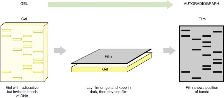
Once the sample is labeled, the labeled tissue section is put next to an X-ray film or nuclear emulsion to make an autoradiograph.
When beta particles interact with the silver ions in the photographic emulsion, which is made of silver bromide crystals in a gelatin matrix, they turn on Ag+ ions.
During development, the activated Ag+ ions are turned into Ag(s), leaving grains of Ag(s) to mark the path of the beta particles.
Autoradiography can be a simple method, but it does require being careful with radioactive materials to keep everyone safe.
Operators should take the right steps to protect themselves from harmful radiation.
Tip: Turn on the caption button if you need it. Choose “automatic translation” in the settings button, if you are not familiar with the English language (or Indian accent). You may need to click on the language of the video first before your favorite language becomes available for translation.
Applications of Autoradiography
Autoradiography is a method that can be used in many different kinds of biological research.
This article will give an overview of some of the most important uses of autoradiography, such as DNA fingerprinting and genetic analysis, as well as how it is used to study metabolism, pharmacokinetics, and neurobiology.
DNA Fingerprinting and Genetic Analysis
Autoradiography is a key part of DNA fingerprinting, which has changed forensic science, paternity disputes, and immigration cases.
It works by using probes to bind to specific DNA sequences and then using different detection methods, such as autoradiography, to see the bound probes.
After gel electrophoresis and the development of a film that was left in contact with the gel, Jeffreys got an autoradiogram with a number of dark bands.
These dark bands were sections of DNA that had a sequence that matched the probe.
Autoradiography can also be used to analyze the amount of radiation in DNA array autoradiographs, which are used in paternity cases as genetic markers.
The technique lets researchers see specific pieces of DNA on an X-ray film. This gives them important information about when and where cells form.
https://www.sciencedirect.com/topics/agricultural-and-biological-sciences/autoradiography
Metabolism and Pharmacokinetics
Autoradiography has been used to study the metabolism of both plants and animals by keeping track of the activity of radioactive isotopes in organic compounds that have been put into the tissue.
It can be used to find out where a radioactive substance is in a tissue or cell after it has been put into a metabolic pathway, bound to a receptor or enzyme, or hybridized to a nucleic acid.
Autoradiography can also be used to find out where a radiolabeled drug is in the body and how well it binds to a receptor.
For example, autoradiography is often used to study how nucleic acids mix and to measure the amount of radiolabeled drugs in serum for pharmacokinetic studies.
Neurobiology
Autoradiography and radio-labeled compounds are used in neurobiological research to study nerve pathways and receptors.
By seeing how radioactively labeled compounds are distributed in the brain, researchers can learn more about the mechanisms behind normal and abnormal brain function.
Protein Localization
Autoradiography can also be used to find out where proteins are in cells. In this case, a radioactive isotope is added to a protein, and the labeled protein is put into the cells.
The cells are then treated and put on a film or plate for photography. This makes an image of where the labeled protein is in the cell. This lets scientists study how different proteins in cells work and how they are controlled.
Receptor Localization
Autoradiography can also be used to find receptors inside cells and study how they work. In this case, a radioactive ligand is used to mark the receptors. The cells are then processed and put on a film or plate for photography.
This makes a picture of where the labeled receptors are inside the cells. This lets researchers study where receptors are and what role they play in cell signaling and other things that cells do.
Radioligand Binding Assays
In radioligand binding assays, autoradiography is often used to look at how ligands and receptors work together. In this application, a radioactive ligand is mixed with cells or tissues, and autoradiography is used to measure how well the ligand binds to the receptors.
This lets researchers study the speed and strength of the interactions between ligands and receptors and find potential drugs or other compounds that could change these interactions.
Alternatives to Autoradiography
Autoradiography is a common way to find out if something has radioactivity in it.
But there are a number of other ways to find and measure radioactive isotopes, and some of them have better sensitivity and resolution.
Imaging Plate Autoradiography
Imaging Plate (IP) autoradiography is a simple, non-destructive way to analyze samples
It can take pictures of large areas in two dimensions and has low detection limits for actinides and other radioactive nuclides.
The radiation given off by the radioactive isotope is caught by a storage phosphor screen, which is then read by a scanner and turned into a digital image.
Scanning Electron Microscopy (SEM)

Scanning electron microscopy (SEM) is a method that uses an electron beam to make high-resolution pictures of microscopic objects.
SEM can also be used to look at how radioisotopes are spread out in samples.
The sample is covered with a material that conducts electricity, and the electron beam scans over the sample's surface to make images with high resolution and good contrast.
https://en.wikipedia.org/wiki/Scanning_electron_microscope
Secondary Ion Mass Spectrometry (SIMS)
Secondary Ion Mass Spectrometry (SIMS) is a method that can be used to find and take pictures of isotopes that are smaller than a micron.
For this method, a beam of high-energy ions is fired at the sample, which makes secondary ions come out.
The mass spectrometer is then used to look at these ions to find out where and how many isotopes there are in the sample.
Phosphor Screen Autoradiography
Using the 14C-PMMA method, Phosphor Screen autoradiography is a technique that uses a radioactive isotope to figure out how porous something is and what its pores look like.
For this method, PMMA resin is poured around a sample, which is then exposed to a radioactive isotope.
The sample is then pictured using a phosphor screen, which picks up the sample's radioactive emissions.
Other Alternatives
Besides these methods, the following are also common alternatives to autoradiography:
- Liquid scintillation counting is a method for detecting and measuring low levels of beta and alpha emitting isotopes that is both sensitive and quantitative.
- Gamma counting is used to find and measure the amount of gamma emitters in different types of samples.
Labeling and Detecting Proteins
Autoradiography is a type of imaging that uses radioactive sources that are already present in the sample, such as radiolabeled proteins.
During protein synthesis, radioactive isotopes like 35S-methionine, 3H-leucine, or 14C-amino acids can be added to the protein of interest
This makes it possible to use autoradiography to find and measure labeled proteins.
This method is especially useful for finding proteins that are not very common or for looking at how proteins change after they are made.
Through co-immunoprecipitation and overlay assays, autoradiography can also be used to find out how proteins interact with each other.
Labeling and Detecting DNA
By adding radioactive isotopes like sulfur-35 (35S), hydrogen-3 (3H), carbon-14 (14C), iodine-125 (125I), and phosphorus-32 (32P) to the DNA molecule, autoradiography can also be used to mark and find DNA.
For example, 32P and 35S can be added to nucleosides like N15- or deoxythymidine triphosphate (dTTP), which can then be used to label DNA molecules.
In proliferation assays, you can also use 3H-thymidine or thymidine that has been labeled with 14C.
Autoradiography can also be used to find out how 32P-radiolabeled oligonucleotides are being used to fix DNA.
Radiation Safety and Research Setting
Autoradiography is a method used in biological research to see radiolabeled proteins, DNA, and other parts in a sample and figure out how much of each there is.
It involves putting a piece of labeled tissue next to a piece of photographic film or emulsion. This makes an autoradiograph.
Autoradiographs can be looked at through a microscope to find out where silver grains are, like on the inside or outside of cells or organelles.
When using radioactive materials in research, there are a number of ways to stay safe.
- Designating and labeling areas where radioactive materials will be used.
- You can not eat, drink, or smoke in the lab.
- Using spill trays and a covering that soaks up liquid.
- Using fume hoods when working with materials that could catch fire.
- Putting on personal protective gear like lab coats, gloves, and safety glasses.
- Keeping an eye on surfaces and cleaning them after use.
- Putting radioactive waste in trash cans the right way, as required by law.
Direct autoradiography with film is limited in sensitivity by the inefficient transfer of emission energy of radionuclides.
Conclusion
As we finish learning about autoradiography, one thing is clear: there is no denying the power of radioactivity in biological research.
Autoradiography has helped us learn a lot about the natural world, from the time scientists found it over a hundred years ago to the present, when it is used in fields like genetics and neuroscience.
But it is important to remember that when you have a lot of power, you also have a lot of responsibility.
Autoradiography is a powerful way to find out about things, but it must be used carefully and with care to avoid the risks of radiation exposure.
As an engineer, you have the rare chance to work at the cutting edge of science, using new methods like autoradiography to learn more about the world around us.
By keeping an eye on safety and pushing the limits of what is possible, you can help make sure that this amazing technology will continue to lead to new discoveries for many years to come.
So go forth, explore, and discover the amazing world of autoradiography – the possibilities are endless!
Share on…
